The annual School of Chemical Sciences (SCS) Science Image Challenge is held for researchers to engage the public with images that are educational and inspirational. This competition is open to undergraduate students, graduate students, postdoctoral associates/fellows, and staff. Posters from the challenge are also presented in the Willard Airport. To learn more about the SCS Science Image challenge and to see winners from previous years, visit the SCS SIC webpage.
Below are the 2023 challenge winners and finalists:
Main Category winner
Rose in the Flask
Xiaolin Liu, Moore’s Lab, Department of Chemistry
Some roses grow from flowerbeds, and others appear in chemistry labs. This rose-like hue and pattern formed at the bottom of a flask as dichloromethane solvent evaporated from an aromatic compound sample. The polycyclic aromatic hydrocarbons show useful electronic properties and also imparts color.
Main Category Finalists
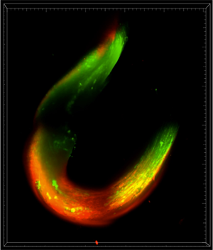 Muscle-bot
Kai-Yu Huang, Kong lab, Department of Chemical & Biomolecular Engineering
The fluorescent image represented a 3D engineered muscle bundle composed of mouse skeletal muscle. The image was acquired with the Lightsheet Z1 system in Carl R. Woese Institute for Genomic Biology.
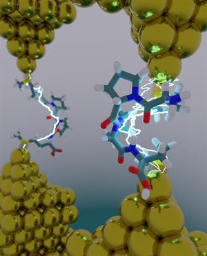 Do Peptides Dream of Electric Sheep?
Moeen Meigooni, Tajkhorshid Lab & Rajarshi Samajdar, Schroeder Group, Center for Biophysics and Quantitative Biology, Departments Biochemistry, Chemistry, & Chemical & Biomolecular Engineering, Beckman Institute
An atomistic model of a molecular break junction: an oligopeptide is caught between two gold electrodes, forming a single-molecule circuit. Using a combined experimental- computational approach, we show that secondary structure plays an important role in determining electron transport in peptides. Image rendered using VMD and Blender
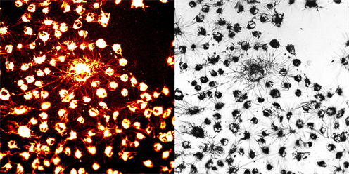 Arachnid Constellations in Cellular Alchemy
Suritra Bandyopadhyay, Chan Lab, Department of Chemistry
What may appear as a spider web or fireworks illuminating the sky are indeed formazan crystals formed within living cells through the action of mitochondrial enzymes. While this process is employed to evaluate the cellular toxicity of chemicals, it showcases the intersection of biology and art through microscopy
 Ugly duckling poly-donuts
Wangsuk Oh, Xiao Su lab, Department of Chemical & Biomolecular Engineering
Polymer electrolyte complexes can form particles with diverse shapes like bread dough. The electron microscopy image shows an uncommon donut-shaped (or toroidal) particle surrounded by spherical nanoparticles formed when drying the polymer electrolyte mixtures. Image obtained at the Materials Research Laboratory Facilities
 Golden Neckless
Xiaolin Liu, Moore lab, Department of Chemistry
The “golden necklace” pattern was formed because of trapped air and water pockets during frontal ring-opening metathesis polymerization, an energy-efficient technique for fabricating high-quality thermosets and composites used in engineering applications
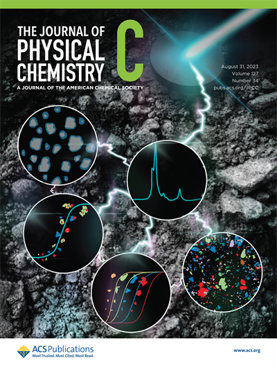 Cover Art Category Winner
Yating Feng & Krishna C. Polavaram, Garg Lab, Departments of Civil & Environmental Engineering & Department of Chemistry
Using Raman imaging, for the first time, we report phase-specific particle size distributions and shape characteristics for each phase in a set of 10 commercially available cements. By integrating these physical characteristics with chemical abundance, we establish a new parameter (CSQ) that can predict the 72 h cumulative heat of hydration (R2 = 0.86). This finding underscores the significance of phase-specific particle size distributions in predicting the performance of cementitious systems.